Looking for design inspiration?   Browse our curated collections!

Previous Image

|

Next Image
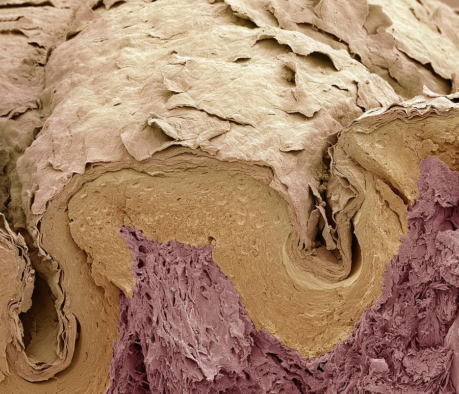
Scanning Electron Micrograph Photograph - Finger skin, SEM by Science Photo Library

Finger skin, SEM is a photograph by Science Photo Library which was uploaded on March 4th, 2014.

More from Science Photo Library

View Profile

Dendritic cell, SEM by Science Photo Library
Mesenchymal stem cell, SEM #1 by Science Photo Library
T Lymphocytes And Cancer Cell, Sem by Steve Gschmeissner
Bone Cross-section #4 by Science Photo Library
Macrophage Engulfing Tb Bacteria by
Dividing Pollen Cell #4 by Professor T. Naguro
Scanning Electron Microscopy by Pascal Goetgheluck/science Photo Library
Scanning Electron Microscope by Karl Gaff / Science Photo Library
Scanning Electron Microscope by Science Photo Library
Scanning Electron Microscope Column Set-up by Elena Hartley/elabarts/science Photo Library
Scanning Electron Microscope Column Set-up #1 by Elena Hartley/elabarts/science Photo Library
Scanning Electron Microscope Control Panel by Elena Hartley/elabarts/science Photo Library
Scanning Electron Microscopy #3 by Pascal Goetgheluck/science Photo Library
Scanning Electron Microscope Control Panel #1 by Elena Hartley/elabarts/science Photo Library
Dendritic Cell #6 by Steve Gschmeissner
Bone Cross-section by Science Photo Library
Fibroblast Cell #4 by Steve Gschmeissner/science Photo Library
Bronchial Epithelium #1 by Steve Gschmeissner
Lavender Leaf by Karl Gaff
Neuron With Amyloid Beta Peptide by Linnea Rundgren/linear Imaging/science Photo Library

Comments

There are no comments for Finger skin, SEM.   Click here to post the first comment.

Recently Viewed

Finger skin, SEM by Science Photo Library
Previous Image Next Image