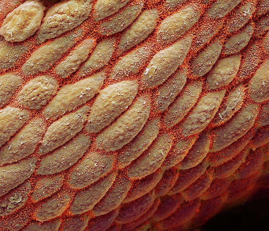
Colon #1 is a photograph by Steve Gschmeissner which was uploaded on July 20th, 2016.
Colon #1
Colon. Coloured scanning electron micrograph (SEM) of the surface of the human colon. Absorptive cells predominate with characteristic microvilli to... more
Title
Colon #1
Artist
Steve Gschmeissner
Medium
Photograph
Description
Colon. Coloured scanning electron micrograph (SEM) of the surface of the human colon. Absorptive cells predominate with characteristic microvilli to increase the surface area and remove water and any remaining nutrients from the already digested food. Magnification: x2300 when printed at 10 centimetres wide.
Uploaded
July 20th, 2016
More from Steve Gschmeissner
Comments
There are no comments for Colon #1. Click here to post the first comment.


