Looking for design inspiration?   Browse our curated collections!

Previous Image

|

Next Image
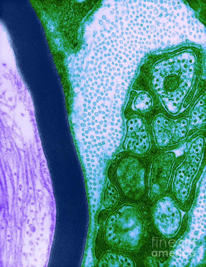
Microscopy Photograph - Nerve Cell, Tem #1 by David M. Phillips

Nerve Cell, Tem #1 is a photograph by David M. Phillips which was uploaded on June 30th, 2014.

More from This Artist

View Profile

Diatoms #6 by M I Walker
Skin Cancer, Bowens Disease, Lm by Garry DeLong
Neuron, Tem #1 by Science Source
Mitochondrion, Tem #2 by Keith R Porter
Neuron, Tem by Science Source
Streptococcus Pyogenes Bacteria Sem #3 by Science Source
Anthrax bacteria SEM by Eye Of Science and Photo Researchers
Ketamine Crystals by M. I. Walker
Peronospora Parasitica #1 by Biophoto Associates and Photo Researchers
Naegleria Fowleri, Lm by Science Source
Motor Neuron From Spinal Cord by M I Walker
Mature Graafian Follicle, Mouse Ovary by M. I. Walker
Hyaline Cartilage #1 by M. I. Walker
Hyacinth Root Tip Cells by Omikron
Skin, Lm #9 by Alvin Telser
Light Micrograph Of Tilia Stem by Lauren Piedmont
Flu Virus Tem by Science Source
Lm Of Skeletal Muscle #1 by Eric V. Grave
Human Sperm, Dic by M. I. Walker
Diaptomus by M. I. Walker

Comments

There are no comments for Nerve Cell, Tem #1.   Click here to post the first comment.

Recently Viewed

Nerve Cell, Tem #1 by David M. Phillips
Previous Image Next Image