Looking for design inspiration?   Browse our curated collections!

Previous Image

|

Next Image
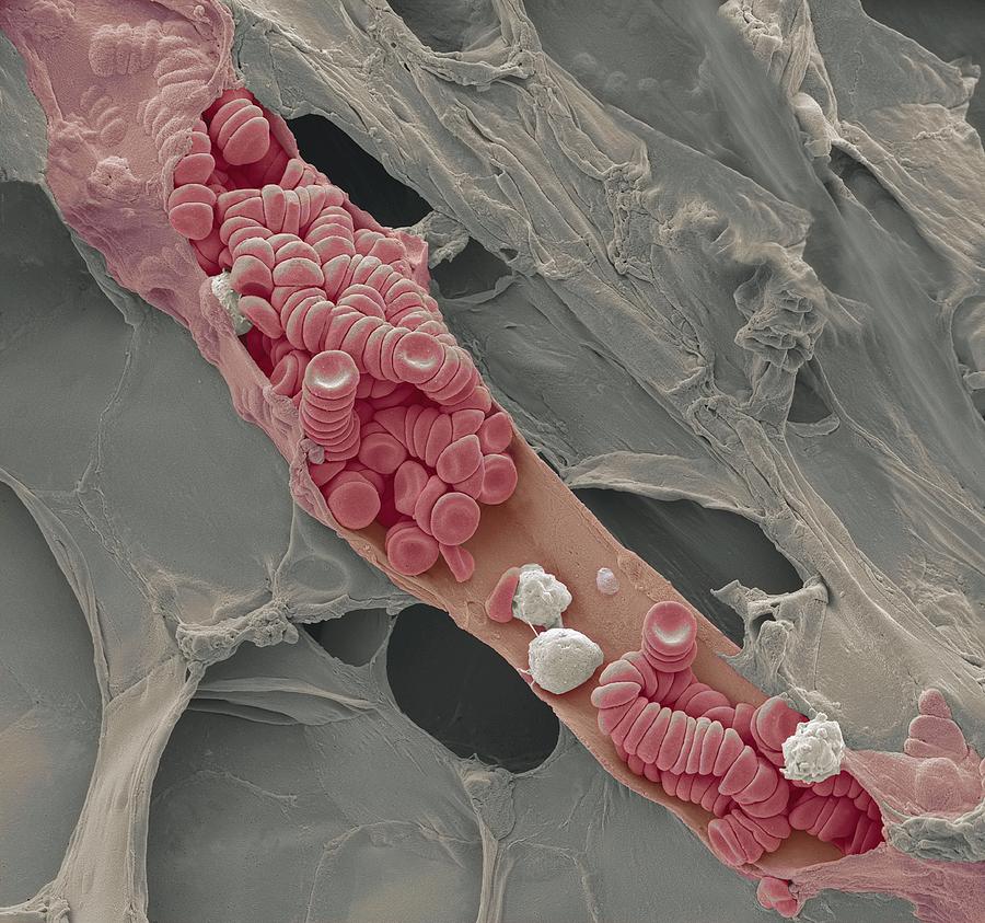
Electron Microscope Photograph - Ruptured Venule, Sem #1 by Steve Gschmeissner

Ruptured Venule, Sem #1 is a photograph by Steve Gschmeissner which was uploaded on May 12th, 2013.

More from Steve Gschmeissner

View Profile

Dendritic cell, SEM by Science Photo Library
Mesenchymal stem cell, SEM #1 by Science Photo Library
T Lymphocytes And Cancer Cell, Sem by Steve Gschmeissner
Bone Cross-section #4 by Science Photo Library
Macrophage Engulfing Tb Bacteria by
Scanning Electron Microscope by Karl Gaff / Science Photo Library
Scanning Electron Microscope by Science Photo Library
Transmission Electron Microscope by Mauro Fermariello/science Photo Library
Scanning Electron Microscope by Colin Cuthbert/science Photo Library
Scanning Electron Microscope #4 by Colin Cuthbert/science Photo Library
Scanning Electron Microscope #5 by Colin Cuthbert/science Photo Library
Liver Cells by Medimage/science Photo Library
Transmission Electron Microscopy by Crown Copyright/health & Safety Laboratory Science Photo Library
Transmission Electron Microscopy by Jim West
Transmission Electron Microscopy #1 by Crown Copyright/health & Safety Laboratory Science Photo Library
Scanning Electron Microscopy by Crown Copyright/health & Safety Laboratory Science Photo Library
Transmission Electron Microscopy #2 by Crown Copyright/health & Safety Laboratory Science Photo Library
Transmission Electron Microscope Cables by Ibm Research
Electron Microscope by Gustoimages/science Photo Library
Electron Microscopy by Gustoimages/science Photo Library

Comments

There are no comments for Ruptured Venule, Sem #1.   Click here to post the first comment.

Recently Viewed

Ruptured Venule, Sem #1 by Steve Gschmeissner
Previous Image Next Image