Purkinje Cells, Light Micrograph is a photograph by Steve Gschmeissner which was uploaded on May 6th, 2013.
Purkinje Cells, Light Micrograph
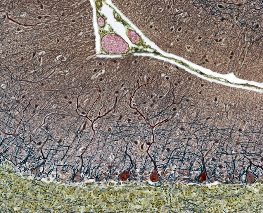
Purkinje cells. Light micrograph of a section through the cerebellum, which has been treated with silver stains, showing pukinje cells (dark brown)... more
Title
Purkinje Cells, Light Micrograph
Artist
Steve Gschmeissner
Medium
Photograph
Description
Purkinje cells. Light micrograph of a section through the cerebellum, which has been treated with silver stains, showing pukinje cells (dark brown) and their dendritic processes. Magnification x250 when printed at 10 centimetres wide.
Uploaded
May 6th, 2013
More from Steve Gschmeissner
Comments
There are no comments for Purkinje Cells, Light Micrograph. Click here to post the first comment.


